Detection-Guided Kidney Abnormality Segmentation
Tumors and cysts are two major kidney abnormalities which can lead to cancer if left undetected and untreated timely. The current diagnostic process depend on computed tomography (CT) scan screening which is time-consuming and specialist-dependent. This leads to fatigue for radiologists and doctors. As a result, diagnostic errors increase. To enhance the diagnostic process, we are developing an AI-based automated method for kidney abnormality detection.
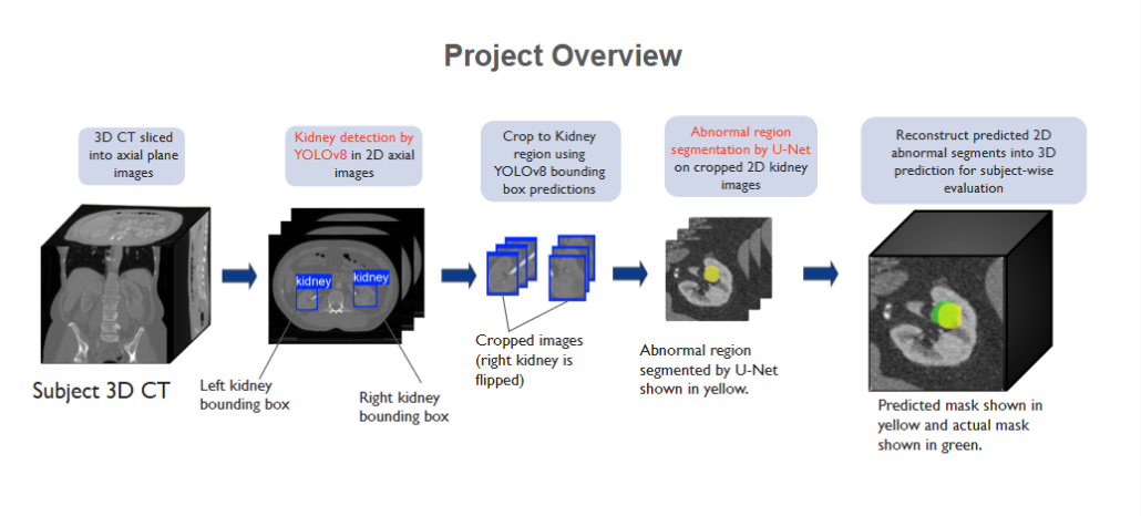
The first step in this method is kidney detection using the YOLOv8 model. This is conducted on 2D sliced images extracted from 3D CT. The detected kidney regions are then cropped to reduce the background area. After that, abnormal region segmentation is conducted using the U-Net segmentation model. The abnormal region consists of either cyst or tumor or both. This produces a mask of abnormal region for each 2D slice. The 2D mask slices are combined to construct a 3D mask of the abnormal region.
This study aims to automate the detection process of kidney tumor and cyst, offering a faster and enhanced approach to assist the doctors and radiologists in diagnostic process.
Relevant publications:
1. J. Faruk, S. B. Alam, S. S. T. Elma, S. Wasi, R. Rahman and S. Kobashi, “Kidney Abnormality Detection Using Segmentation-Guided Classification on Computed Tomography Images,” 2024 International Conference on Machine Learning and Cybernetics (ICMLC), Miyazaki, Japan, 2024, pp. 414-419, doi: 10.1109/ICMLC63072.2024.10935113.
2. S. Wasi, S. B. Alam, R. Rahman, M. A. Amin and S. Kobashi, “Kidney Tumor Recognition from Abdominal CT Images using Transfer Learning,” 2023 IEEE 53rd International Symposium on Multiple Valued Logic (ISMVL), Matsue, Japan, 2023, pp. 54-58, doi: 10.1109/ISMVL57333.2023.00021